Monoclonal antibodies targeting calcitonin gene-related peptide show good efficacy as preventive migraine therapy however, their site of action is not fully understood. In a special update session at #MTIS2020, Jacob Edvinsson, University of Copenhagen, Denmark highlighted recent research that reveals insights into the role of CGRP in neuronal cross-talk in the trigeminal ganglion, which plays a central role in migraine pathophysiology.
CGRP-mediated cross-talk in the trigeminal ganglion
As monoclonal antibodies (mAbs) targeting calcitonin gene-related peptide (CGRP) are not able to cross the blood-brain barrier, the trigeminovascular system is proposed as a possible site of action of CGRP mAbs because of the peripheral location of the trigeminal nerves. Cross-talk of signals between trigeminal ganglion neurons and neighboring neurons and satellite glial cells involves intracellular mechanisms that can modulate mediators of sensory information, such as neuropeptides including CGRP, receptors and neurotrophic factors.1 Drugs that modulate cross-talk in the trigeminal ganglion can therefore modulate both peripheral and central sensitization.2
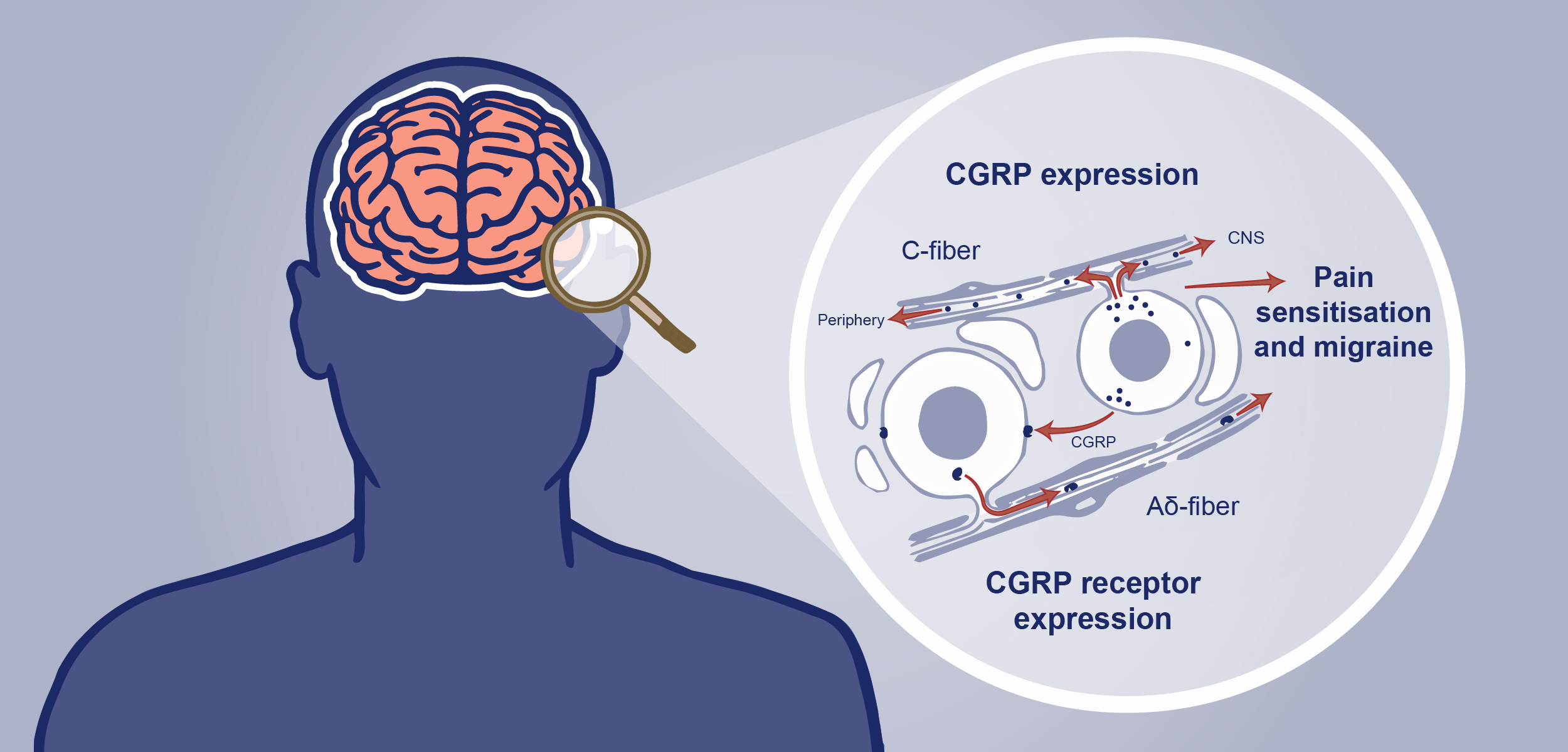
The persistent modulatory effects of CGRP through C-fiber and Aδ-fibers cross-talk may lead to central pain sensitization and chronification of migraine
Jacob Edvinsson outlined recent imaging studies that are helping to better understand the role of CGRP in migraine. There are two populations of neurons identified in the trigeminal ganglion, C-fibers expressing CGRP and Aδ-fibers that express the CGRP receptor.3 C-fibers are unmyelinated fibers that contain pearl-like synaptic structures –boutons – and release CGRP in the trigeminal ganglion.
Imaging reveals that CGRP-containing C-fiber boutons align with the node of Ranvier in Aδ-fibres.3 The relevance of this alignment is not yet known but could suggest a new site of action for C-fiber signaling in the trigeminal ganglion. CGRP may act on adjacent myelinated axons via CGRP receptor localization in the node of Ranvier and the paranodes (axon-axon signaling). Adjacent to the CGRP receptor localization in the node of Ranvier, immunoreactivity to protein kinase A (PKA; a kinase stimulated by cAMP) was identified, providing a structural possibility for modifying conduction activity within the Aδ-fibers. Thus, the neuromodulatory CGRP signaling pathway may affect membrane potentials (sensitization) through PKA-mediated activation of sodium/potassium voltage channels. The persistent modulatory effects of CGRP through C-fiber and Aδ-fibers cross-talk may lead to central pain sensitization and chronification of migraine.4
The close proximity of CGRP-containing C-fibers and Aδ-fibers containing CGRP receptor elements in the trigeminal ganglion suggests a site of action for CGRP antibodies to alleviate migraine
The observed close relationship between CGRP containing C-fibers and the Aδ-fibers containing the CGRP-receptor elements, suggests a point of axon-axon interaction for the released CGRP and a site of action for gepants and the novel CGRP mAbs to alleviate migraine.3
Our correspondent’s highlights from the symposium are meant as a fair representation of the scientific content presented. The views and opinions expressed on this page do not necessarily reflect those of Lundbeck.